Austin Lim, PhD
Cart 0

Academic Publications

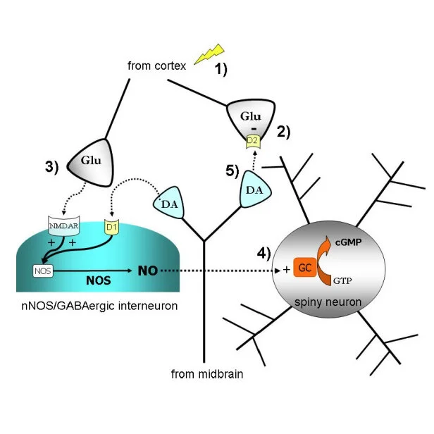
My primary interest in neurobiology is to understand the circuitry of a brain region called the striatum. The striatum is important for everyday functions such as motor learning, decision making, and motivation. Improper neuronal signaling in the striatum is related to a wide variety of disorders, including Parkinson's disease, addiction, schizophrenia, and OCD. Getting a clearer picture of the way parts of the brain communicate with the striatum will help us develop therapies for these disorders.

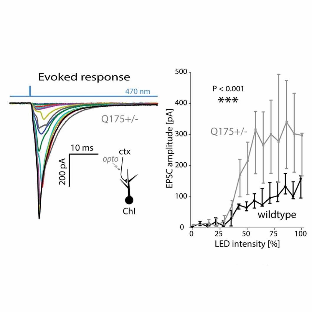
I use electrophysiological techniques in combination with optogenetics and pharmacology to understand the cells in the striatum. The majority of my research uses patch-clamp electrophysiology (whole cell, cell attached, and perforated patch) in an ex vivo slice preparation. Using this technique, I am able to study the way striatal cells communicate.


Huntington's disease

is a rare genetic disorder characterized by frequent uncontrollable movements, cognitive difficulties, and emotional dysregulation. It is often diagnosed in middle age, and is lethal within a decade or two. It has a very strong genetic component associated with the onset and severity of the disease. Although there is an FDA approved therapy for the symptoms, the exact nature of the changes in the brain are not fully characterized. My research with the Surmeier group at Northwestern University has focused largely on understanding the changes in the striatum related to Huntington's disease.

 
Lim SAO and Surmeier DJ (2021) Enhanced GABAergic Inhibition of Cholinergic Interneurons in the zQ175+/− Mouse Model of Huntington's Disease. Front. Syst. Neurosci. 14:626412. doi: 10.3389/fnsys.2020.626412

Lim SAO and Surmeier DJ (2021) Enhanced GABAergic Inhibition of Cholinergic Interneurons in the zQ175+/− Mouse Model of Huntington's Disease. Front. Syst. Neurosci. 14:626412. doi: 10.3389/fnsys.2020.626412

ENHANCED GABAERGIC INHIBITION OF CHOLINERGIC INTERNEURONS IN THE ZQ175+/− MOUSE MODEL OF HUNTINGTON'S DISEASE

Huntington's disease (HD) is an autosomal dominant neurodegenerative disorder that initially manifests itself in the striatum. How intrastriatal circuitry is altered by the disease is poorly understood. To help fill this gap, the circuitry linking spiny projection neurons (SPNs) to cholinergic interneurons (ChIs) was examined using electrophysiological and optogenetic approaches in ex vivo brain slices from wildtype mice and zQ175+/− models of HD. These studies revealed a severalfold enhancement of GABAergic inhibition of ChIs mediated by collaterals of indirect pathway SPNs (iSPNs), but not direct pathway SPNs (dSPNs). This cell-specific alteration in synaptic transmission appeared in parallel with the emergence of motor symptoms in the zQ175+/− model. The adaptation had a presynaptic locus, as it was accompanied by a reduction in paired-pulse ratio but not in the postsynaptic response to GABA. The alterations in striatal GABAergic signaling disrupted spontaneous ChI activity, potentially contributing to the network dysfunction underlying the hyperkinetic phase of HD.


Tanimura A, Lim SAO, Aceves Buendia JJ, Goldberg JA and Surmeier DJ (2016) Cholinergic Interneurons Amplify Corticostriatal Synaptic Responses in the Q175 Model of Huntington’s Disease. Front. Syst. Neurosci. 10:102. doi: 10.3389/fnsys.2016.00102

Tanimura A, Lim SAO, Aceves Buendia JJ, Goldberg JA and Surmeier DJ (2016) Cholinergic Interneurons Amplify Corticostriatal Synaptic Responses in the Q175 Model of Huntington’s Disease. Front. Syst. Neurosci. 10:102. doi: 10.3389/fnsys.2016.00102

CHOLINERGIC INTERNEURONS AMPLIFY CORTICOSTRIATAL SYNAPTIC RESPONSES IN THE Q175 MODEL OF HUNTINGTON'S DISEASE

The synaptic properties of ChIs were examined using optogenetic approaches in the Q175 mouse model of HD. In ex vivo brain slices, synaptic facilitation at thalamostriatal synapses onto ChIs was reduced in Q175 mice. The alteration in thalamostriatal transmission was paralleled by an increased response to optogenetic stimulation of cortical axons, enabling these inputs to more readily induce burst-pause patterns of activity in ChIs. This adaptation was dependent upon amplification of cortically evoked responses by a post-synaptic upregulation of voltage-dependent Na+ channels. This upregulation also led to an increased ability of somatic spikes to invade ChI dendrites. However, there was not an alteration in the basal pacemaking rate of ChIs, possibly due to increased availability of Kv4 channels. Thus, there is a functional “re-wiring” of the striatal networks in Q175 mice, which results in greater cortical control of phasic ChI activity, which is widely thought to shape the impact of salient stimuli on striatal action selection.


Levodopa-induced dyskinesia

is a debilitating motor condition that results following the treatment of Parkinson's disease with dopamine replacement therapy. Patients experiencing LID often exhibit uncontrolled movments (chorea) and painful or debilitating muscle contractions (dystonia). It is estimated that up to 80% of patients with Parkinson's disease will experience these symptoms after 5-10 years of treatment. My work with the McGehee lab in collaboration with the Kang lab at the University of Chicago has expanded our understanding of striatal dysfunction in levodopa-induced dyskinesia. 

 
https://www.sciencedirect.com/science/article/pii/S0969996115000157

https://www.sciencedirect.com/science/article/pii/S0969996115000157

Enhanced histamine H2 excitation of striatal cholinergic interneurons in L-DOPA-induced dyskinesia.

Our previous studies indicate enhanced excitability of striatal cholinergic interneurons in mice expressing LID and reduction of LID when ChIs are selectively ablated. Recent gene expression analysis indicates that stimulatory H2 histamine receptors are preferentially expressed on ChIs at high levels in the striatum, and we tested whether a change in H2 receptor function might contribute to the elevated excitability in LID. Using two different mouse models of PD (6-hydroxydopamine lesion and Pitx3ak/ak mutation), we chronically treated the animals with either vehicle or L-DOPA to induce dyskinesia. Electrophysiological recordings indicate that histamine H2 receptor-mediated excitation of striatal ChIs is enhanced in mice expressing LID. Additionally, H2 receptor blockade by systemic administration of famotidine decreases behavioral LID expression in dyskinetic animals. These findings suggest that ChIs undergo a pathological change in LID with respect to histaminergic neurotransmission. The hypercholinergic striatum associated with LID may be dampened by inhibition of H2 histaminergic neurotransmission. 


https://www.pnas.org/content/108/2/840.full

https://www.pnas.org/content/108/2/840.full

Enhanced striatal cholinergic neuronal activity mediates L-DOPA-induced dyskinesia in parkinsonian mice.

Previous investigations have noted changes in striatal medium spiny neurons, including abnormal activation of extracellular signal-regulated kinase1/2 (ERK). Using two PD models, the traditional 6-hydroxydopamine toxic lesion and a genetic model with nigrostriatal dopaminergic deficits, we found that acute dopamine challenge induces ERK activation in medium spiny neurons in denervated striatum. After repeated L-DOPA treatment, however, ERK activation diminishes in medium spiny neurons and increases in striatal cholinergic interneurons. ERK activation leads to enhanced basal firing rate and stronger excitatory responses to dopamine in striatal cholinergic neurons. Pharmacological blockers of ERK activation inhibit L-DOPA–induced changes in ERK phosphorylation, neuronal excitability, and the behavioral manifestation of LID. In addition, a muscarinic receptor antagonist reduces LID. These data indicate that increased dopamine sensitivity of striatal cholinergic neurons contributes to the expression of LID, which suggests novel therapeutic targets for LID.


Striatal circuitry

is complex in organization, and has not yet been fully characterized. Compared to structures such as the cortex with a laminar organization, the striatum consists of a jumble of cells with few discrete boundaries. While working with the Surmeier lab at Northwestern University, the McGehee lab at the University of Chicago, and the West lab at Rosalind Franklin University, I have furthered our understanding of the organization of the striatum and the various neurochemical signals that alter striatal function.

 
Tanimura, A., Pancani, T., Lim, S.A.O., Tubert, C., Melendez, A.E., Shen, W. and Surmeier, D.J. (2018), Striatal cholinergic interneurons and Parkinson's disease. Eur J Neurosci, 47: 1148-1158. https://doi.org/10.1111/ejn.13638

Tanimura, A., Pancani, T., Lim, S.A.O., Tubert, C., Melendez, A.E., Shen, W. and Surmeier, D.J. (2018), Striatal cholinergic interneurons and Parkinson's disease. Eur J Neurosci, 47: 1148-1158. https://doi.org/10.1111/ejn.13638

Striatal cholinergic interneurons and Parkinson's disease

Giant, aspiny cholinergic interneurons (ChIs) have long been known to be key nodes in the striatal circuitry controlling goal‐directed actions and habits. In recent years, new experimental approaches, like optogenetics and monosynaptic rabies virus mapping, have expanded our understanding of how ChIs contribute to the striatal activity underlying action selection and the interplay of dopaminergic and cholinergic signaling. These approaches also have begun to reveal how ChI function is distorted in disease states affecting the basal ganglia, like Parkinson's disease (PD). This review gives a brief overview of our current understanding of the functional role played by ChIs in striatal physiology and how this changes in PD. The translational implications of these discoveries, as well as the gaps that remain to be bridged, are discussed as well.


Lim SAO, Kang UJ and McGehee DS (2014) Striatal cholinergic interneuron regulation and circuit effects. Front. Synaptic Neurosci. 6:22. doi: 10.3389/fnsyn.2014.00022

Lim SAO, Kang UJ and McGehee DS (2014) Striatal cholinergic interneuron regulation and circuit effects. Front. Synaptic Neurosci. 6:22. doi: 10.3389/fnsyn.2014.00022

Striatal cholinergic interneuron regulation and circuit effects

The striatum plays a central role in motor control and motor learning. Appropriate responses to environmental stimuli, including pursuit of reward or avoidance of aversive experience all require functional striatal circuits. These pathways integrate synaptic inputs from limbic and cortical regions including sensory, motor and motivational information to ultimately connect intention to action. Although many neurotransmitters participate in striatal circuitry, one critically important player is acetylcholine (ACh). Relative to other brain areas, the striatum contains exceptionally high levels of ACh, the enzymes that catalyze its synthesis and breakdown, as well as both nicotinic and muscarinic receptor types that mediate its postsynaptic effects. The principal source of striatal ACh is the cholinergic interneuron (ChI), which comprises only about 1–2% of all striatal cells yet sends dense arbors of projections throughout the striatum. This review summarizes recent advances in our understanding of the factors affecting the excitability of these neurons through acute effects and long term changes in their synaptic inputs. In addition, we discuss the physiological effects of ACh in the striatum, and how changes in ACh levels may contribute to disease states during striatal dysfunction.


Ondracek, J.M., Dec, A., Hoque, K.E., Lim, S.A.O., Rasouli, G., Indorkar, R.P., Linardakis, J., Klika, B., Mukherji, S.J., Burnazi, M., Threlfell, S., Sammut, S. and West, A.R. (2008), Feed‐forward excitation of striatal neuron activity by frontal c…

Ondracek, J.M., Dec, A., Hoque, K.E., Lim, S.A.O., Rasouli, G., Indorkar, R.P., Linardakis, J., Klika, B., Mukherji, S.J., Burnazi, M., Threlfell, S., Sammut, S. and West, A.R. (2008), Feed‐forward excitation of striatal neuron activity by frontal cortical activation of nitric oxide signaling in vivo. European Journal of Neuroscience, 27: 1739-1754. https://doi.org/10.1111/j.1460-9568.2008.06157.x

Feed-forward excitation of striatal neuron activity by frontal cortical activation of nitric oxide signaling in vivo

The gaseous neurotransmitter nitric oxide plays an important role in the modulation of corticostriatal synaptic transmission. This study examined the impact of frontal cortex stimulation on striatal nitric oxide efflux and neuron activity in urethane‐anesthetized rats using amperometric microsensor and single‐unit extracellular recordings, respectively. Systemic administration of the neuronal nitric oxide synthase inhibitor 7‐nitroindazole decreased spontaneous spike activity without affecting activity evoked by single‐pulse stimulation of the ipsilateral cortex. Train (30 Hz) stimulation of the contralateral frontal cortex transiently increased nitric oxide efflux in a robust and reproducible manner. Evoked nitric oxide efflux was attenuated by systemic administration of 7‐nitroindazole and the non‐selective nitric oxide synthase inhibitor NG‐nitro‐l‐arginine methyl ester. Train stimulation of the contralateral cortex, in a manner identical to that used to evoke nitric oxide efflux, had variable effects on spike activity assessed during the train stimulation trial, but induced a short‐term depression of cortically evoked activity in the first post‐train stimulation trial. Interestingly, 7‐nitroindazole potently decreased cortically evoked activity recorded during the train stimulation trial. Moreover, the short‐term depression of spike activity induced by train stimulation was enhanced following pretreatment with 7‐nitroindazole and attenuated after systemic administration of the dopamine D2 receptor antagonist eticlopride. These results demonstrate that robust activation of frontal cortical afferents in the intact animal activates a powerful nitric oxide‐mediated feed‐forward excitation which partially offsets concurrent D2 receptor‐mediated short‐term inhibitory influences on striatal neuron activity. Thus, nitric oxide signaling is likely to play an important role in the integration of corticostriatal sensorimotor information in striatal networks.